What is involved in a Retina Exam?
Sometimes called ophthalmoscopy or funduscopy. Performing these tests allows Dr. Zdenek to see the back of your eye; your retina, the optic disk and the underlying layer of blood vessels that nourish the retina (choroid).

Dr. Zdenek may use one or more of these techniques to view the back of your eye:
Direct Exam
Indirect Exam
Retinal Imaging
- Direct exam. Your eye doctor uses an ophthalmoscope to shine a beam of light through your pupil to see the back of the eye. Sometimes eyedrops aren't necessary to dilate your eyes before this exam.
- Indirect exam. During this exam, you might lie down, recline in a chair or sit up. Your eye doctor examines the inside of the eye with the aid of a condensing lens and a bright light mounted on his or her forehead. This exam lets your doctor see the retina and other structures inside your eye in great detail and in three dimensions.
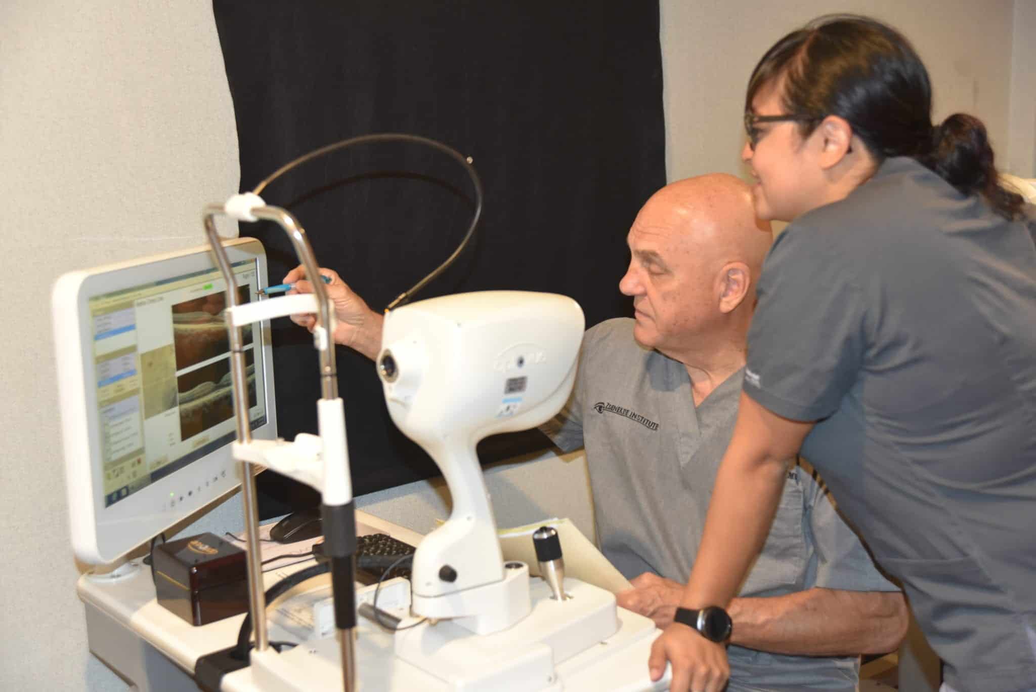
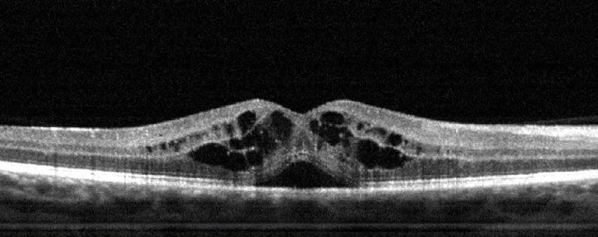
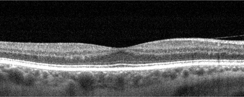
- Retinal Imaging. OCT (Optical Coherence Tomography) is used to capture an image of the retina.
Why get a Retina Exam?
A retinal exam is used in the diagnosis and treatment of Diabetes, Macular degeneration, Glaucoma, and many other diseases. A Retinal exam doesn’t replace your annual eye exam but adds another layer of precision to it.
Finding retinal disorders as early as possible is critical to potentially preventing serious disease progressio.


What Does Your Retina Look Like?
How healthy is your retina?

